Burst fracture radiology
Federal government websites often end in. The site is secure.
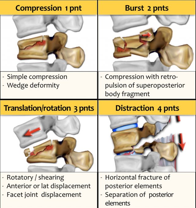
Burst fractures are a type of compression fracture related to high-energy axial loading spinal trauma that results in disruption of a vertebral body endplate and the posterior vertebral body cortex. Retropulsion of posterior cortex fragments into the spinal canal is frequently included in the definition. However, some authors, including the popular AO spine classification system, define a burst fracture as any axial compression fracture involving an endplate and the posterior cortex regardless of retropulsion 6. They usually present as back pain and or lower limbs neurologic deficits in the clinical scenario of trauma. Two-level burst fractures are much less common than single-level burst fractures 2. Burst fractures involve the posterior wall of the vertebral body can be described as incomplete one endplate or complete both endplates 5. See: AO spine classification of thoracolumbar injuries.
Burst fracture radiology
Fifty percent of TL fractures are unstable and can result in significant anatomic injury and deformity 4. Clinical assessment of patients with TL fractures is often challenging and, as a result, diagnostic imaging usually plays an essential role in their exact diagnosis and appropriate management 6. The aim of this article is to review the role of different imaging methods in studying TL fractures, emphasizing the role of the radiologist in classifying and quantifying the severity of these fractures. Radiographs are the adequate starting modality for patients who have sustained a low-energy trauma. AP and lateral views are usually performed. Both projections are useful in assessing vertebral height and the presence of fracture lines. The AP view allows the measurement of the interpedicular distance, which is increased in burst fractures, and the interspinous distance, which is increased in posterior ligamentous complex PLC injuries. These values can be reported as millimeters or as a percentage relative to adjacent normal levels. Regarding interspinous distance, variations of up to 7 mm are considered as normal 7. On lateral radiographs the two main parameters to be measured are vertebral height loss and kyphotic deformity 6 Figure 2.
The teaching point is: pay careful attention to little pieces of bone. MusculoskeletalSpine.
George M. Bridgeforth and Mark Nolden A year-old man falls 30 ft down an elevator shaft without losing consciousness. Burst fractures are traumatic spinal injuries that involve vertebral breakage. They may occur as a result of cervical injuries from diving accidents or from rollover accidents in which the driver was not wearing a seat belt. The mechanism of injury involves an axial load impact against a rock or a shallow river or against the roof of a vehicle as it rolls over.
Single lateral view of the thoracolumbar junction demonstrates a burst fracture of what appears to be L1. CT confirms the presence of a burst fracture with retropulsion of the upper half of the vertebra and significant loss of height particularly anteriorly. Burst fracture with retropulsed fragment, which narrows the central canal. No abnormal spinal cord signal. Updating… Please wait. Unable to process the form. Check for errors and try again.
Burst fracture radiology
Federal government websites often end in. The site is secure. Ankara Cad. Burst fractures can occur with different radiological images after high energy. We aimed to simplify radiological staging of burst fractures. Eighty patients whom exposed spinal trauma and had burst fracture were evaluated concerning age, sex, fracture segment, neurological deficit, secondary organ injury and radiological changes that occurred. We performed a new classification in burst fractures at radiological images. According to this classification system, secondary organ injury and neurological deficit can be an indicator of energy exposure. If energy is high, the clinical status will be worse.
Hallmark cards
First look at the first CT-images and decide what is going on. After several months kyphotic deformity increased D. Case creation learning pathway. Radiology Assistant Information. Case 6 Look at the images. PCL integrity can change treatment to conservative therapy or to minimally invasive surgery 4. Calcifications Differential of Breast Calcifications. A B1 is a monosegmental osseous injury with damage of the posterior tension band; B B2 fracture with ligamentous posterior tension band injury; C B2 fracture with bony posterior tension band injury; D B3 fracture is an anterior tension band injury. At the time the article was created Jeremy Jones had no recorded disclosures. These changes as an indicator of energy that is exposed can also give a hint for secondary organ injury and neurological deficit. METHODS: Eighty patients whom exposed spinal trauma and had burst fracture were evaluated concerning age, sex, fracture segment, neurological deficit, secondary organ injury and radiological changes that occurred. There is a comminuted burst 3 column fracture involving the L1 vertebra, including a large retropulsed fragment causing significant stenosis of the central canal. The bone fragments in the canal were proportional to and neurological deficit secondary organ injury in our study. Always check for bilateral calcaneal injuries from falls. American journal of roentgenology.
Clinical Presentation The patient is a year-old female who states that at approximately p.
A posterior spondylodesis was performed. However the distraction is the most important finding, i. Here a typical case of translation. On lateral radiographs the two main parameters to be measured are vertebral height loss and kyphotic deformity 6 Figure 2. Bridgeforth and Mark Nolden A year-old man falls 30 ft down an elevator shaft without losing consciousness. The aim of this study is to create a simpler radiological classification in the burst fractures and to present their relation to secondary injuries. Sign Up. PLC is used as a reference point while classifying the burst fractures. Bone marrow edema in several vertebral bodies, either due to contusion or fracture. Open in a separate window. On the axial images we see: Transverse process fractures A rib fracture These are typical findings in translation-rotation fractures. Usually, one piece of bone fragment moves on to the spinal canal.


Now all became clear to me, I thank for the necessary information.