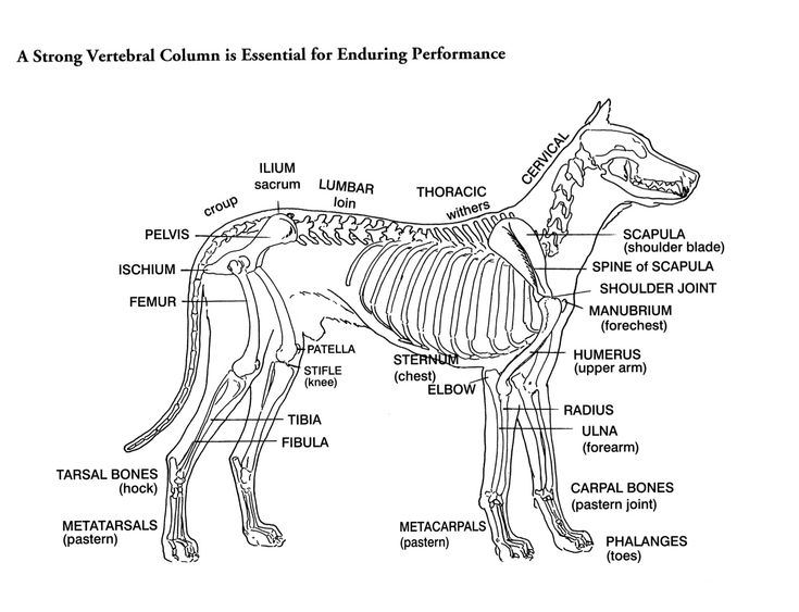
Diagram of a dog skeleton
Allowing for variations in tail length, the canine skeleton consists of an average of individual bones. Bones are complex, rigid, living organs that have diagram of a dog skeleton own supply of blood vessels and nerves. The function of the skeleton is to provide a solid framework for the body and protect internal organs and structures. Each bone moves alongside the contraction and relaxation of muscles, therefore they all have multiple, significant points and structures for muscle attachment.
A — Cervical or Neck Bones 7 in number. B — Dorsal or Thoracic Bones 13 in number, each bearing a rib. C — Lumbar Bones 7 in number. D — Sacral Bones 3 in number. E — Caudal or Tail Bones 20 to 23 in number. Przemek Maksim. If you use on your website or in your publication my images, you are obliged to give following details: - "Author:Przemek Maksim, -graphic name.
Diagram of a dog skeleton
.
These individual bones allow the flexibility and movement of the ankle as well as shock absorption in jumping and weight bearing. Extension of these vertebrae allows the head to be held high and facing the direction of movement whilst on the other hand, allowing forward flexion to reach the ground for sniffing and eating.
.
The dog skeleton anatomy consists of bones, cartilages, and ligaments. You will find two different parts of the dog skeleton — axial and appendicular. Here, I will show you all the bones from the axial and appendicular skeleton with their special osteological features. Again, I will provide more labeled diagrams for each dog skeleton bone. This article will provide a clear conception of the dog paw and foot skeleton anatomy. In addition, I will try to solve the common inquiries on the dog bone anatomy at the end of the article. So, if you are interested to learn the basics of the dog skeleton bones and differentiate them from the other skeletons like goat or horse, you may continue the article till the end. The bones of the dog skeleton anatomy serve to support and protect the visceral organs. Again, all the bones of the dog skeleton provide lavers for muscular action.
Diagram of a dog skeleton
Dog anatomy is not very difficult to understand if a labeled diagram is present to provide a graphic illustration of the same. That is exactly what you will find in this DogAppy article. It provides information about a dog's skeletal, reproductive, internal, and external anatomy, along with accompanying labeled diagrams. After mating, dogs experience something called a copulatory tie, wherein they remain in the coital position. The male dog dismounts the female at this time. The dogs can remain in this position from a few minutes to an hour, and it is recommended not to try and separate them as it can cause injury to their organs. When the pups are born, they have all the bones, muscles, and tendons that an adult dog has.
Starbucks coffee gear coupon code
These individual bones allow the flexibility and movement of the ankle as well as shock absorption in jumping and weight bearing. The most important functions of the ribcage is to protect the lungs, heart and other internal organs of the thorax. The following pages on the English Wikipedia use this file pages on other projects are not listed :. Quality image This image has been assessed using the Quality image guidelines and is considered a Quality image. Carpals- There are a total of 7 carpal bones aligned in two rows parallel to each other forming the wrist. The proximal row consists of the talus and calcaneus forms the hock. Head Skull- A bony structure that supports the structures of the face such as the eyes and provides a protective cavity for the brain. E — Caudal or Tail Bones 20 to 23 in number. Skeleton of a dog diagram. The more uniform shape and longer size means that the lumber area of the spine is commonly the largest in the whole spinal column. As the hind leg extends and flexes, the patella glides up and down within a groove forming the front of the knee joint, thereby protecting the joint. Creative Commons Attribution-ShareAlike 3.
This modules of vet-Anatomy provides a basic foundation in animal anatomy for students of veterinary medicine. This veterinary anatomical atlas includes selected labeling structures to help student to understand and discover animal anatomy skeleton, bones, muscles, joints, viscera, respiratory system, cardiovascular system.
If you use on your website or in your publication my images, you are obliged to give following details: - "Author:Przemek Maksim, -graphic name. Skip to content Allowing for variations in tail length, the canine skeleton consists of an average of individual bones. Email Required Name Required Website. Permission Reusing this file. It is in fact the only mobile bone of the facial skeleton. E — Caudal or Tail Bones 20 to 23 in number. Width The scapulae allows the shoulder to flex, extend, rotate, abduct and adduct. Quality image This image has been assessed using the Quality image guidelines and is considered a Quality image. The function of the skeleton is to provide a solid framework for the body and protect internal organs and structures. The proximal row consists of the radial, ulnar and accessory carpal bones then the distal row consists of carpal bones one to four. Phalanges- The phalanges consist of 4 proximal, 4 middle and 5 4 on Hindlimb distal bones. Share this: Twitter Facebook. This file contains additional information, probably added from the digital camera or scanner used to create or digitize it.


0 thoughts on “Diagram of a dog skeleton”