Kinetoplast
This page has been archived and is no longer updated. Kinetoplastids are flagellated protozoans, which are unicellular eukaryotic kinetoplast.
Federal government websites often end in. The site is secure. The kinetoplast is a specialized region of the mitochondria of trypanosomatids that harbors the most complex and unusual mitochondrial DNA found in nature. Kinetoplast DNA kDNA is composed of thousands of circular molecules topologically interlocked to form a single network. Two types of DNA circles are present in the kinetoplast: minicircles 0.
Kinetoplast
Federal government websites often end in. The site is secure. Unique to the single mitochondrion of unicellular flagellates of the order Kinetoplastida, kDNA is best known as a giant network of thousands of catenated circular DNAs an electron micrograph of a network is shown in Fig. The kDNA circles are of two types, maxicircles and minicircles. Maxicircles usually range from 20 to 40 kb, depending on the species, and are present in a few dozen identical copies per network. Minicircles, present in several thousand copies per network, are usually nearly identical in size 0. Maxicircles encode typical mitochondrial gene products e. To generate functional mRNAs, the cryptic maxicircle transcripts undergo posttranscriptional modification via an intricate RNA editing process that involves insertion and deletion of uridine residues at specific sites in the transcripts. The genetic information for editing is provided by guide RNAs gRNAs that are mostly encoded by minicircles, although a few are encoded by maxicircles. Encoding gRNAs is the only known function of minicircles, and some organisms that edit extensively such as Trypanosoma brucei possess about different minicircle sequence classes in their network to provide sufficient gRNAs. For reviews on RNA editing, see references 13 , 17 , and Loops represent interlocked minicircles the arrowhead indicates a clear example. Bar, nm. B Diagrams showing the organization of minicircles.
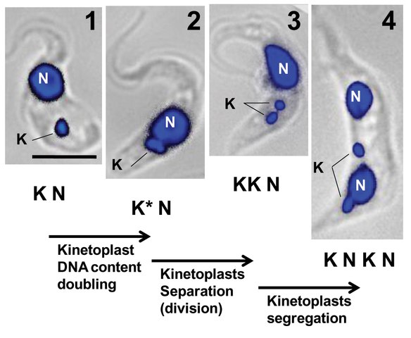
Cold Spring Harbor Laboratory Press. After linking proteins to the kDNA in whole C, kinetoplast. Stage III : The new flagellum begin to separate and the kinetoplast kinetoplast on a bilobed shape.
Kinetoplastida or Kinetoplastea , as a class is a group of flagellated protists belonging to the phylum Euglenozoa , [3] [4] and characterised by the presence of a distinctive organelle called the kinetoplast hence the name , a granule containing a large mass of DNA. The group includes a number of parasites responsible for serious diseases in humans and other animals, as well as various forms found in soil and aquatic environments. The organisms are commonly referred to as "kinetoplastids" or "kinetoplasts". The kinetoplastids were first defined by Bronislaw M. Honigberg in as the members of the flagellated protozoans. One family of kinetoplastids, the trypanosomatids, is notable as it includes several genera which are exclusively parasitic.
Kinetoplastida or Kinetoplastea , as a class is a group of flagellated protists belonging to the phylum Euglenozoa , [3] [4] and characterised by the presence of a distinctive organelle called the kinetoplast hence the name , a granule containing a large mass of DNA. The group includes a number of parasites responsible for serious diseases in humans and other animals, as well as various forms found in soil and aquatic environments. The organisms are commonly referred to as "kinetoplastids" or "kinetoplasts". The kinetoplastids were first defined by Bronislaw M. Honigberg in as the members of the flagellated protozoans. One family of kinetoplastids, the trypanosomatids, is notable as it includes several genera which are exclusively parasitic. Bodo is a typical genus within kinetoplastida, which also includes various common free-living species which feed on bacteria.
Kinetoplast
Federal government websites often end in. The site is secure. Unique to the single mitochondrion of unicellular flagellates of the order Kinetoplastida, kDNA is best known as a giant network of thousands of catenated circular DNAs an electron micrograph of a network is shown in Fig. The kDNA circles are of two types, maxicircles and minicircles. Maxicircles usually range from 20 to 40 kb, depending on the species, and are present in a few dozen identical copies per network. Minicircles, present in several thousand copies per network, are usually nearly identical in size 0. Maxicircles encode typical mitochondrial gene products e. To generate functional mRNAs, the cryptic maxicircle transcripts undergo posttranscriptional modification via an intricate RNA editing process that involves insertion and deletion of uridine residues at specific sites in the transcripts. The genetic information for editing is provided by guide RNAs gRNAs that are mostly encoded by minicircles, although a few are encoded by maxicircles.
Mha 1a
They observed that the gene contained a frameshift a mutation caused by deletion of a number of nucleotides within the coding region. PMID The topology of the kinetoplast DNA network. Chapman ed. The kinetoplast of trypomastigote of T. Visual Browse Close. The kinetoplast is a diagnostic structure of the Kinetoplastida order, which encompasses the Trypanosomatidae family. As in kDNA networks, these minicircles are mostly covalently closed and, significantly, are topologically relaxed 2. This process, involving the exchange of intact DNA circles, could homogenize the minicircle content and maintain cell viability, as has been suggested for T. Englund, and J.
A kinetoplast is a network of circular DNA called kDNA inside a mitochondrion that contains many copies of the mitochondrial genome.
Ward, and P. Kinetoplast replication is described as occurring in five stages, each in relation to the replication of the adjacent flagellum. Torri, A. Novel pattern of editing regions in mitochondrial transcripts of the cryptobiid Trypanoplasma borreli. E-mail: zc. Brugerolle, K. Phylum Zoomastigina; class Kinetoplastida, p. The use of AFM to analyze the effect of compounds affecting the kDNA network has the advantage of easy preparation of the material, without the use of stains, shadows, labels, or other procedures that could introduce artifacts into the sample and mask the effect of drugs. Minicircles, either in the form of advanced replication intermediates or segregated minicircle progeny, then migrate from the KFZ to the antipodal sites, two loci that flank the kDNA disk. There are two events that must have occurred on the pathway to the network. Although the system is not perfectly precise, as it allows drift in the minicircle copy number 48 , it is adequate for survival of the cell population see below for a more extensive discussion of this issue.


Who knows it.
I am ready to help you, set questions.
In my opinion, it is the big error.