Lipoma on pancreas
Regret for the inconvenience: we are taking measures to prevent fraudulent form submissions by extractors and page crawlers. Received: October 27, Published: November 27,
Federal government websites often end in. The site is secure. Correspondence to: Dr. Lipomas of the pancreas are very rare. There are fewer than 25 reported cases of lipoma originating from the pancreas.
Lipoma on pancreas
Pancreatic lipomas are thought to be very rare. Lipomas are usually easy to identify on imaging, particularly via computed tomography CT. Here, we present a case of pancreatic lipoma in a year-old female. She was asymptomatic and had no medical history of note. Finally, the patient underwent a pancreaticoduodenectomy. Histologically, mature adipocytes were noted in the bulk of the tumor. Accordingly, the pathologic diagnosis of the pancreatic neoplasm was lipoma. To our knowledge, this case is the first example of a suspected well-differentiated liposarcoma that was actually a pancreatic lipoma. We also highlight the radiological features distinguishing a pancreatic lipoma from a pancreatic liposarcoma and briefly review the literature. Pancreatic lipomas show no obvious gender bias and most commonly occur in the head of the pancreas, of which the maximum diameters are often less than 5 cm, and small, asymptomatic non-compressed lipomas require follow-up only. Surgical excision should be considered when the tumor has compressed important tissues or is difficult to distinguish from a liposarcoma, the choice of surgery depends on the intraoperative presentation. Core tip: Pancreatic lipomas are rare, especially the huge ones. Lipomas are usually easily identified on imaging, particularly via computed tomography.
We discuss and highlight the radiological features distinguishing a pancreatic lipoma from other fatty lesions of the pancreas and pancreatic liposarcoma and provide a brief review of the literature. A lipoma on pancreas woman with a past medical history of dyslipidemia and cholelithiaisis was admitted to our institution for right hypochondrium pain and epigastric discomfort and vomiting of 2 wk duration, lipoma on pancreas.
At the time the article was last revised Daniel J Bell had no financial relationships to ineligible companies to disclose. Pancreatic lipomas are uncommon mesenchymal tumors of the pancreas. Rarely symptomatic, they are most often detected incidentally on cross-sectional imaging for another purpose. If they do cause symptoms, it will typically be those related to regional mass effect from the mass. Pancreatic lipomas are composed of mature fat cells with thin internal fibrous septa.
Hence, localizing the tumor site can guide the healthcare provider to arrive at a probable diagnosis. The specific risk factors for Lipoma of Pancreas are unknown or unidentified. Note: It is important to note that an individual diagnosed with cancer of the pancreas may not have any of the above-mentioned risk factors. It is important to note that having a risk factor does not mean that one will get the condition. A risk factor increases ones chances of getting a condition compared to an individual without the risk factors. Some risk factors are more important than others. Also, not having a risk factor does not mean that an individual will not get the condition. It is always important to discuss the effect of risk factors with your healthcare provider.
Lipoma on pancreas
Federal government websites often end in. The site is secure. Recent studies have shown a significant increase in the utilization of computed tomography CT scans in the emergency department for a broad spectrum of conditions. This had a significant impact on the identification of patients with serious pathologies in a timely manner. However, the overutilization of computed tomography scans leads to increased identification of incidental findings.
Stones to kilograms
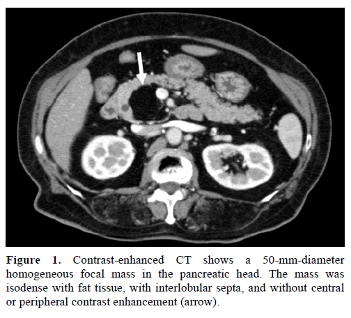
We feared that complete removal would increase the risk of injury to the pancreatic duct and superior mesenteric vein, which might trigger a major intraoperative hemorrhage and a postoperative pancreatic fistula that could erode the superior mesenteric vein and cause a massive hemorrhage or other complications. In summary, lipomas of the pancreas are very rare. Loading more images Focal fatty masses of the pancreas. Morgan's current disclosures. Pancreatic lipomas Lipoma of the pancreas Lipomas of the pancreas. Most contain macroscopic fat. MRI is very useful and shows almost total replacement of pancreatic parenchyma by fatty tissue. Unlike other pancreatic tumors, it can be confirmed on CT or MRI imaging and does not require invasive histopathological examination to establish a definite diagnosis. The borders were indistinct and a few fibroreticular septa were evident within the lesion. Pancreatic lipoma seems to exhibit no gender bias, is usually diagnosed via CT or other imaging methods, and most commonly occurs in the head of the pancreas. T2 magnetic resonance image. Publishing Process of This Article. Well-differentiated liposarcoma presents as high signal intensity on T1-weighted T1W images, intermediate signal intensity on T2-weighted T2W images and drop-out signal intensity on fat-suppressed MR images.
At the time the article was last revised Daniel J Bell had no financial relationships to ineligible companies to disclose. Pancreatic lipomas are uncommon mesenchymal tumors of the pancreas.
Federal government websites often end in. Copy Download. The natural history of pancreatic lipoma: Does it need observation. Teratoma is also rare in the pancreas and is diagnosed when a variegated density lesion is present or both calcification and fat are present in the lesion. Pancreatic lipomas show no obvious gender bias and most commonly occur in the head of the pancreas, of which the maximum diameters are often less than 5 cm, and small, asymptomatic non-compressed lipomas require follow-up only. Butler et al. This cross sectional image reveals a 6. Updating… Please wait. Write to the Help Desk. This cross sectional image reveals a homogenous fatty containing lesion in the head of pancreas. Most contain macroscopic fat. As present, imaging techniques are very accurate, and in most cases, there is no need for histopathological confirmation of pancreatic lipoma. Abdominal Imaging. Of these rare tumors, fat-originating tumors lipomas and liposarcomas are the rarest.


Yes cannot be!
It is remarkable, rather useful message
I apologise, but you could not give little bit more information.