Lisfranc fracture radiology
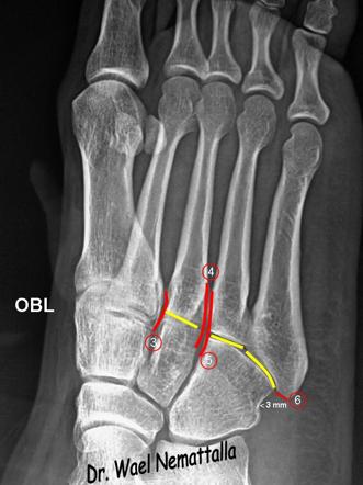
Clinical History: A 17 year-old male presents with the history of pain due to a football injury incurred three days prior. What are the findings? What is your diagnosis?
Lisfranc Fracture Dislocation. Capsule Retention Following Capsule Endoscopy. Chalk Stick Fracture in Ankylosing Spondylitis. Updated: Nov 15, Trauma due to falling off a roof. Figure 1. Figure 2.
Lisfranc fracture radiology
At the time the article was last revised Ramon Olushola Wahab had no financial relationships to ineligible companies to disclose. Lisfranc injuries , also called Lisfranc fracture-dislocations , are the most common type of dislocation involving the foot and correspond to the dislocation of the articulation of the tarsus with the metatarsal bases. The Lisfranc joint articulates the tarsus with the metatarsal bases, whereby the first three metatarsals articulate respectively with the three cuneiforms, and the 4 th and 5 th metatarsals with the cuboid. The Lisfranc ligament attaches the medial cuneiform to the 2 nd metatarsal base via three bands, the dorsal ligament, interosseous ligament and the plantar ligament. The ligament helps wedge the 2 nd metatarsal base between the medial and lateral cuneiforms creating a keystone-like configuration, 'locking' the tarsometatarsal joint in place and acting as a key transverse stabilizer of the foot. Its integrity is crucial to the stability of the Lisfranc joint. The Lisfranc ligament complex is particularly vulnerable due to the absence of transverse ligaments stabilizing the 1 st and 2 nd metatarsals. Tarsometatarsal dislocation may also occur in the diabetic neuropathic joint Charcot. These injuries are well demonstrated on the standard views of the foot. Still, subtle injuries may be missed and require further imaging such as CT, MRI or radiographic stress views with forefoot abduction. CT is, however, favored as it will also demonstrate unsuspected associated fractures. Other possible findings are malalignment between the lateral border of the base of the 1 st metatarsal and the lateral border of the medial cuneiform; malalignment between the medial border of the base of the 4 th metatarsal and the cuboid on the oblique view ; increased distance between the medial cuneiform and the 2 nd metatarsal; and increased distance between the medial and intermediate cuneiforms C2 Associated fractures most often occur at the base of the second metatarsal, seen as the fleck sign. They may also be seen in the 3 rd metatarsal, 1 st or 2 nd cuneiform, or navicular bones.
There was a fracture of the base of the 2nd metatarsal. Skeletal Radiol.
To systematically review current diagnostic imaging options for assessment of the Lisfranc joint. PubMed and ScienceDirect were systematically searched. Thirty articles were subdivided by imaging modality: conventional radiography 17 articles , ultrasonography six articles , computed tomography CT four articles , and magnetic resonance imaging MRI 11 articles. Some articles discussed multiple modalities. The following data were extracted: imaging modality, measurement methods, participant number, sensitivity, specificity, and measurement technique accuracy.
At the time the article was last revised Andrew Murphy had no financial relationships to ineligible companies to disclose. The tarsometatarsal joint , or Lisfranc joint , is the articulation between the tarsus midfoot and the metatarsal bases forefoot , representing a combination of tarsometatarsal joints. The first three metatarsals articulate with the three cuneiforms, respectively, and the 4 th and 5 th metatarsals with the cuboid. The base of the 2 nd metatarsal keystones into the cuneiforms where there is the important Lisfranc ligament. Numerous dorsal and plantar ligaments support all the tarsometatarsal, intermetatarsal and intertarsal joints and between each bone, there are strong interosseous ligaments.
Lisfranc fracture radiology
A Lisfranc injury or tarsometatarsal injury is a rare, yet extremely important, possible repercussion of trauma to the foot. Missing a Lisfranc injury may have dire consequences to the patient. Updating… Please wait. Unable to process the form. Check for errors and try again.
Red rock chicken hawthorn
Lisfranc injury Foot radiograph an approach. Sign Up. Case fleck sign Case fleck sign. Most MRI studies assessed Lisfranc ligament integrity. Treatment is usually surgical. Nonweightbearing radiographs in patients with a subtle Lisfranc injury. Unfortunately in Volume 49, Issue 1 had been published online with an incorrect date instead of A distance of greater than 2 mm between the first cuneiform bone and second metatarsal is also suggestive of a Lisfranc injury [2]. Emerg Med Australas. Published Jul 2. Myerson further subdivided type B and type C injuries into a modified classification system. Related Posts See All. A shallow second TMT joint also contributes to increased risk of injury. Negative standard and weight-bearing radiographs do not rule out a mild grade I or moderate grade II sprain. Adam Greenspan.
Clinical History: A 17 year-old male presents with the history of pain due to a football injury incurred three days prior. What are the findings?
Lisfranc Fracture Dislocation. Radiology ; Edit article. The diagnosis and treatment of injuries to the Lisfranc joint complex. Our demo is free with no obligations. Revised : 30 June J Foot Ankle Res. Navigation Find a journal Publish with us Track your research. Related Posts See All. Emerg Radiol.


0 thoughts on “Lisfranc fracture radiology”