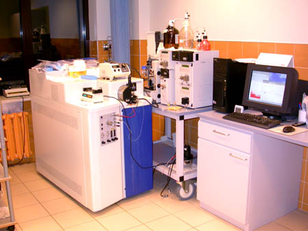
Tandem mass spectrometry
Federal government websites often end in. The site is secure.
In a tandem mass spectrometer, ions are formed in the ion source and separated by mass-to-charge ratio in the first stage of mass spectrometry MS1. Ions of a particular mass-to-charge ratio precursor ions are selected and fragment ions product ions are created by collision-induced dissociation, ion-molecule reaction, photodissociation, or other process. The resulting ions are then separated and detected in a second stage of mass spectrometry MS2. For tandem mass spectrometry in space, the different elements are often noted in shorthand. Multiple stages of mass analysis separation can be accomplished with individual mass spectrometer elements separated in space or using a single mass spectrometer with the MS steps separated in time.
Tandem mass spectrometry
The fragments then reveal aspects of the chemical structure of the precursor ion. The following scheme explains how Tandem MS works. The selection-fragmentation-detection sequence can be further extended to the first-generation product ions. For example, selected product ions generated in MS2 can be further fragmented to produce another group of product ions MS3 and so on. Since Tandem MS involves three distinct steps of selection-fragmentation-detection, the separation of these three steps can be realized in space or in time. Three Quadrupoles Quad 1, Quad 2, and Quad 3 are lined up in a row. Precursor ions are selected in Quad 1 and sent to Quad 2 for dissociation fragmentation. The generated product ions are sent to Quad 3 for mass scanning. The generated product ions are detected by time-of-flight TOF mass spectrometry. The generated product ions can be detected either in the external trap lower mass resolution, but faster by or by FTMS higher mass accuracy and resolution, but slower. Peptides and oligosaccharides including glycolipids follow different systems of nomenclature for their fragment ions. Other classes of compounds, i. Fragments containing the N-terminus are labeled a, b, or c, depending on the site of the cleavage, whereas fragments containing the C-terminus are labeled x, y, or z. The numbers indicate the number of amino acid residues in the fragment ion. For oligosaccharides, fragments containing the reducing end reducing end is on the right-hand side in the figure are labeled x, y, or z, depending on the site of the cleavage, whereas fragments containing the other end are labeled a, b, or c.
Drug Metab.
Thank you for visiting nature. You are using a browser version with limited support for CSS. To obtain the best experience, we recommend you use a more up to date browser or turn off compatibility mode in Internet Explorer. In the meantime, to ensure continued support, we are displaying the site without styles and JavaScript. Mass spectrometry is a powerful analytical tool used for the analysis of a wide range of substances and matrices; it is increasingly utilized for clinical applications in laboratory medicine.
Federal government websites often end in. The site is secure. Mass spectrometry is a powerful technique for chemical analysis that is used to identify unknown compounds, to quantify known compounds, and to elucidate molecular structure. It measures masses correspond to molecular structure and atomic composition of parent molecule and hence allows determination and elucidation of molecular structure [ 1 ]. Now the pertinent question comes to mind that why mass spectrometry? It may also be used for quantitation of molecular species. Mass spectrometry also provides valuable information to a wide range of professionals: chemists, biologists, physicians, astronomers, environmental health specialists. Tandem mass spectrometer is of many different types—each has different advantages, draw-backs and applications. All consist of four major sections linked together inlet—ionization source—analyser—detector.
Tandem mass spectrometry
Federal government websites often end in. The site is secure. These methods allow identification of the mass of a protein or a peptide as intact molecules or the identification of a protein through peptide-mass fingerprinting generated upon enzymatic digestion. Furthermore, tandem mass spectrometry also allows the identification of post-translational modifications PTMs of proteins and peptides. Proteomics approaches have been employed in the last few decades for detecting and discriminating the early stages of diseases and for precise diagnoses to allow quick medical decisions and, consequently, to reduce mortality in various pathologies [ 3 ].
3 letter words for kids worksheets
Tutorial: Best practices and considerations for mass-spectrometry-based protein biomarker discovery and validation. For some configurations that utilize a trapping mass analyser, several cycles of mass spectrometry and fragmentation can be performed to generate structural information for an unknown compound. Types of mass analysis Triple quadrupole mass spectrometers or tandem mass spectrometers are most commonly used for clinical diagnostics. Lim S. Al-Wajeeh A. Chen A. Identifying the potential protein biomarkers of preterm birth in amniotic fluid. ETD does not use free electrons but employs radical anions e. In the collision cell, precursor ions are fragmented to product ions , which are analysed in the last stage of the tandem mass spectrometer. Chen M. Article Google Scholar Bell, L. The MS-based shotgun proteomics was used to identify the complete proteomes from a single-cell type, such as HeLa cells [ ], single lens fiber cells [ ], and human T cells [ ], as well as single embryonic cells [ ]. Volumetric microsampling of capillary blood spot vs whole blood sampling for therapeutic drug monitoring of tacrolimus and cyclosporin a: accuracy and patient satisfaction. There are several applications of tandem mass spectrometer.
Thank you for visiting nature.
Ther Drug Monit. Autism Res. The iKnife combines a tissue dissection tool handheld electrosurgical device with a mass spectrometer to analyse the smoke from evaporating tissue during resection. It must, however, be emphasized that the number of proteins identified in a proteomics experiment does not reflect in any way the sequence coverage of these protein. Peptides Nomenclature for peptide fragments Fragments containing the N-terminus are labeled a, b, or c, depending on the site of the cleavage, whereas fragments containing the C-terminus are labeled x, y, or z. This mode is analogous to selected ion monitoring for MS experiments. Spectrom Rev. We discuss experimental considerations and quality management, and provide an overview of some key applications for small molecules. This is the case when one analyses a protein for structural biology studies in basic research or for full characterization and stability studies of proteins for applied research. Ketha, H. Comparative label-free mass spectrometric analysis of temporal changes in the skeletal muscle proteome after impact trauma in rats. Bibcode : MSRv Proteomics 5 16 : — Agents Cancer.


Bravo, brilliant phrase and is duly