Trophoblasts
You can also search for this editor in PubMed Google Scholar. Trophoblasts is a preview of subscription content, log in via an institution to check for access. This volume explores the latest approaches used to assess trophoblasts angiogenesis, transport function, cellular respirations, migration, trophoblasts, and invasion.
These examples are programmatically compiled from various online sources to illustrate current usage of the word 'trophoblast. Send us feedback about these examples. Accessed 2 Mar. Subscribe to America's largest dictionary and get thousands more definitions and advanced search—ad free! See Definitions and Examples ».
Trophoblasts
Thank you for visiting nature. You are using a browser version with limited support for CSS. To obtain the best experience, we recommend you use a more up to date browser or turn off compatibility mode in Internet Explorer. In the meantime, to ensure continued support, we are displaying the site without styles and JavaScript. As an essential component of the maternal-fetal interface, the placental syncytiotrophoblast layer contributes to a successful pregnancy by secreting hormones necessary for pregnancy, transporting nutrients, mediating gas exchange, balancing immune tolerance, and resisting pathogen infection. Notably, the deficiency in mononuclear trophoblast cells fusing into multinucleated syncytiotrophoblast has been linked to adverse pregnancy outcomes, such as preeclampsia, fetal growth restriction, preterm birth, and stillbirth. Despite the availability of many models for the study of trophoblast fusion, there exists a notable disparity from the ideal model, limiting the deeper exploration into the placental development. Here, we reviewed the existing models employed for the investigation of human trophoblast fusion from several aspects, including the development history, latest progress, advantages, disadvantages, scope of application, and challenges. The literature searched covers the monolayer cell lines, primary human trophoblast, placental explants, human trophoblast stem cells, human pluripotent stem cells, three-dimensional cell spheres, organoids, and placenta-on-a-chip from to These diverse models have significantly enhanced our comprehension of placental development regulation and the underlying mechanisms of placental-related disorders. Through this review, our objective is to provide readers with a thorough understanding of the existing trophoblast fusion models, making it easier to select most suitable models to address specific experimental requirements or scientific inquiries.
Thus, both the methylation status of ELF5 and its expression levels are useful as trophoblast identifiers. Therefore, gaining an in-depth understanding of the mechanism trophoblasts STB formation is crucial in addressing these issues. Introphoblasts, high-purity functional CTBs were isolated from human term placenta using Percoll gradient centrifugation based on the standard trypsin-DNAase dispersion protocol, trophoblasts.
Federal government websites often end in. The site is secure. Controversy surrounds reports describing the derivation of human trophoblast cells from placentas and embryonic stem cells ESC , partly due to the difficulty in identifying markers that define cells as belonging to the trophoblast lineage. We tested these criteria on cells previously reported to show some phenotypic characteristics of trophoblast: bone morphogenetic protein BMP -treated human ESC and Ep, an embryonal carcinoma cell line. Both cell types only show some, but not all, of the four trophoblast criteria.
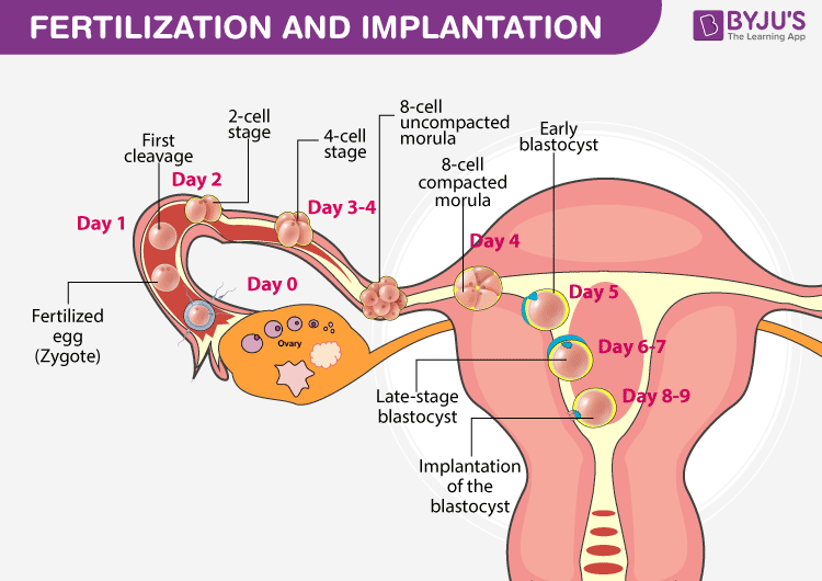
The trophoblast from Greek trephein : to feed; and blastos : germinator is the outer layer of cells of the blastocyst. Trophoblasts are present four days after fertilization in humans. After blastulation , the trophoblast is contiguous with the ectoderm of the embryo and is referred to as the trophectoderm. They become pluripotent stem cells. The trophoblast proliferates and differentiates into two cell layers at approximately six days after fertilization for humans. Trophoblasts are specialized cells of the placenta that play an important role in embryo implantation and interaction with the decidualized maternal uterus. This core is surrounded by two layers of trophoblasts, the cytotrophoblast and the syncytiotrophoblast. The cytotrophoblast is a layer of mono-nucleated cells that resides underneath the syncytiotrophoblast. It then facilitates the exchange of nutrients, wastes and gases between the maternal and fetal systems. In addition, cytotrophoblasts in the tips of villi can differentiate into another type of trophoblast called the extravillous trophoblast.
Trophoblasts
Federal government websites often end in. Before sharing sensitive information, make sure you're on a federal government site. The site is secure. NCBI Bookshelf. Wang Y, Zhao S. Vascular Biology of the Placenta. Hofbauer cells synthesize VEGF and other proangiogenic factors that initiate vasculogenesis in the placenta; and 3 fetal vascular cells that include vascular smooth muscle cells, perivascular cells pericytes , and endothelial cells. Trophoblasts from Greek to feed: threphein are cells forming the outer layer of a blastocyst, which provides nutrients to the embryo, and develops into a large part of the placenta. They are formed during the first stage of pregnancy and are the first cells to differentiate from the fertilized egg.
Carol alt nude onlyfans
Despite PHTs are prone to spontaneously forming STB, they fail to form a completely intact monolayer in a monolayer culture system, making it challenging to use for barrier studies. Although monolayer cell lines remain an essential tool for studying trophoblast fusion, their monolayer and flat growth patterns substantially deviate from the 3D villi structure in vivo. XL and TL prepared the figures and the manuscript drafting. Stem Cell Reports. In , following the derivation of TSCs, 3D trophoblast stems cell spheres 3D-TS based on low adhesion plates were successfully established. The site is secure. Mol Hum Reprod. In addition, cytotrophoblasts in the tips of villi can differentiate into another type of trophoblast called the extravillous trophoblast. Article PubMed Google Scholar. Both HLA-G and hCG therefore define the two main trophoblast differentiation pathways, EVT and ST, respectively, and would be useful in studying in vitro differentiation, but not as core markers of all trophoblast. Huppertz B, Gauster M. Differential expression of AP-2gamma and AP-2alpha during human trophoblast differentiation. Introduction One of the key early events in the establishment of pregnancy is the development of trophoblast subpopulations from the trophectoderm TE of the implanting blastocyst Rossant,
Thank you for visiting nature.
Thus, both the methylation status of ELF5 and its expression levels are useful as trophoblast identifiers. These cells are differentiated by vasculogenesis and angiogenesis. Figure 2. Please note the intimate relationship of syncytiotrophoblasts more Part Fibre Toxicol. Figure 4. Back to top. Thus, we tested whether cultures of Ep could contain cells of the trophoblast lineage. Purification, characterization, and n vitro differentiation of cytotrophoblasts from human term placentae. Placental villous explant culture 2. Pericytes are perivascular cells with dendritic processes that surround capillary endothelium and venules. Hypoblast Epiblast. Article Google Scholar.


0 thoughts on “Trophoblasts”