What is hydatid cyst
Federal government websites often end in. The site is secure. Language: English Turkish. Hydatid disease HD is a unique parasitic disease that primarily affects the liver and is endemic in many parts of the world.
Federal government websites often end in. The site is secure. Hydatid cyst is a parasitic infection that primarily affects the liver but which can be found anywhere in the body. This case involves spontaneous dissemination of hydatid cyst disease, a rare occurrence in the absence of any intervention or trauma. A year-old male patient complained of abdominal pain of 10 years' duration. He was apparently asymptomatic until 10 years ago, at which point he developed abdominal pain and vomiting.
What is hydatid cyst
A hydatid cyst is like a little water balloon in your liver. It contains clear liquid and the young form of a parasite called a dog tape worm or echinococcus. They are very rare in the UK. Most people with hydatid cysts will have got the condition overseas. Often the cysts do not cause any issues. But in very rare cases they can burst and cause a severe allergic reaction. This needs immediate medical help. Back to top. Young dog tape worms live in sheep and other farm animals. Dogs can pick them up if they eat raw meat from an animal that had the condition. Once the tape worms are in a dog they can become adults and lay eggs. Humans can accidently swallow the eggs. For example by kissing a dog. Or not washing your hands properly before touching your mouth after handling dog poo.
If you use automatic language translation services in connection with this site you do so at your own risk. True wall enhancement is seen in infected Type I cysts. Links with this icon indicate that you are leaving the CDC website, what is hydatid cyst.
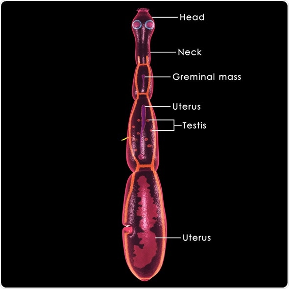
Hydatid disease is caused by infection with a small tapeworm parasite called Echinococcus granulosus. In Australia, most infections are passed between sheep and dogs, although other animals including goats, horses, kangaroos, dingoes and foxes may be involved. The hydatid parasite is carried by dogs in their bowel, without any symptoms of infection. Sheep become infected while grazing in areas contaminated with dog faeces. Dogs become infected by eating the uncooked organs of infected sheep. People become infected by ingesting eating eggs of the parasite, usually when there is hand-to-mouth transfer of eggs in dog faeces.
Federal government websites often end in. The site is secure. Introduction: the hydatic disease, caused by the larvae of Echinococcus granulosus, is a serious disease, potentially lethal, which can be found anywhere in the world, but especially in endemic areas such as the Mediterranean Basin, Australia, New Zealand, North Africa, Eastern Europe, the Balkans, Middle East and South America. The hepatic hydatid cyst therapy is multimodal, including medical, surgical, and, lately, minimally invasive techniques. Materials and methods: 88 patients were diagnosed with liver hydatid cyst at the General Surgery Clinic of the Colentina Hospital in Bucharest where they were admitted from January to July Age, gender, place of origin, year and duration of admission, symptoms and signs at admission, paraclinical serological tests relevant for liver function and E. Conclusions: patients with hepatic hydatid cyst form a heterogeneous group, semiology being poor and unspecific. Among the laboratory examinations, eosinophilia is a sign of concern but is present in less than half of the patients. Imaging findings are the basis for the diagnosis of hepatic hydatid cysts. The hydatic disease is a severe, potentially lethal disease caused by Echinococcus granulosus larvae.
What is hydatid cyst
Most are harmless, but they should be removed when possible because they occasionally may change into malignant growths, become infected, or obstruct a gland. There are four main types of cysts: retention cysts , exudation cysts , embryonic cysts , and parasitic cysts. Baker cyst a swelling on the back of the knee, due to escape of synovial fluid that has become enclosed in a sac of membrane. Bartholin cyst a mucus-filled cyst of a Bartholin gland, usually developing as a consequence of an obstruction of the duct by trauma, infection, epithelial hyperplasia, or congenital atresia or narrowing. See also cystic disease of breast. An example is a branchial cyst. Called also enterocyst and enterocystoma. Called also wen. See also hydatid disease.
Ttd3
In some cases, you might have a general anaesthetic. Figure 2. Most people only get symptoms years after picking them up. Large cysts in the hepatic parenchyma can cause biliary duct dilatation by either compression of a nearby duct by mass effect or by perforation into biliary ducts. Aspiration sclerotherapy might be used if your cyst has got very big and is causing symptoms. CT is superior in detecting gas or air-fluid levels within the cyst Figure Abbreviations: CT, computed tomography; HD, hydatid disease. These HCs are seen in three types: 1 Total and thick continuous calcification ring-like of the cyst wall Figure 5 , 2 total calcification within the cyst matrix and a decrease in cyst size Figure 6 , and 3 curvilinear calcification within the ruptured internal membranes Figure 7. This means there is no parasite growing in them. You can also get hydatid cysts in other parts of your body, including your lungs. However, a simple liver cyst along with a solitary HC as well as polycystic liver disease in the presence of multiple type I HCs can cause diagnostic problems. Heymann, D. Person-to-person or sheep-to-person transmission does not occur. This appearance is not diagnostic for echinococcosis.
Ever since Hippocrates described Hydatid disease, physicians all over the world have encountered it in various organs. Hydatid disease is also referred to as echinococcosis or echinococcal disease. It results from an infection due to a tapeworm of genus Echinococcus.
Introduction Hydatid disease HD is a common parasitic disease produced by the larval stage of the Echinococcus tape-worm. Type I HCs become especially important when they exert a mass effect. Contact Us. But it can give doctors an idea of how your liver is doing. Talk to your doctor about which option will be best for you. This test will look at several things in your blood. And help to rule out more common liver problems. In the peritoneum, they appear as multiple cystic lesions, or with calcification when they are in the inactive or healed stage. There is thick, circumferential calcification at the right-sided cyst. Your doctor will need to look at your scan results to see how big the cysts are and where they are. CT of the thorax Fig. Once the ingested embryos enter the portal circulation, they primarily affect the liver, but can be spread hematogeneously to all organs and tissues except hair. With rare portal vein compression and decreased vascular supply, the involved lobe may show atrophic changes while the other lobe becomes hypertrophic. From to there were between 4 notifications each year.


In my opinion you have gone erroneous by.