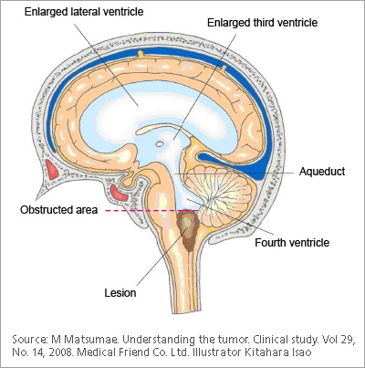
Cerebral aqueduct stenosis
Cerebral aqueduct stenosis Sylvian aqueduct is a narrow channel, about 15 mm long, that connects the third and the fourth ventricle. Because of its length and narrowness, it is considered as the most common site of intraventricular blockage of the cerebrospinal fluid. In this chapter, pathological and etiological findings, specific clinical aspects, neuroradiological appearance, and therapeutic options of hydrocephalus secondary to aqueductal stenosis are exhaustively reviewed, cerebral aqueduct stenosis. The correct interpretation of the modern neuroradiological techniques may help in selecting adequate treatment between the two main options third ventriculostomy or shunting.
Aqueductal stenosis is a narrowing of the aqueduct of Sylvius which blocks the flow of cerebrospinal fluid CSF in the ventricular system. The aqueduct of Sylvius is the channel which connects the third ventricle to the fourth ventricle and is the narrowest part of the CSF pathway with a mean cross-sectional area of 0. This blockage causes ventricle volume to increase because the CSF cannot flow out of the ventricles and cannot be effectively absorbed by the surrounding tissue of the ventricles. Increased volume of the ventricles will result in higher pressure within the ventricles, and cause higher pressure in the cortex from it being pushed into the skull. A person may have aqueductal stenosis for years without any symptoms, and a head trauma , hemorrhage , or infection could suddenly invoke those symptoms and worsen the blockage. Many of the signs and symptoms of aqueductal stenosis are similar to those of hydrocephalus. These typical symptoms include: headache, nausea and vomiting, cognitive difficulty, sleepiness, seizures, balance and gait disturbances, visual abnormalities, and incontinence.
Cerebral aqueduct stenosis
Aqueductal stenosis is a narrowing stenosis of the small connecting duct between the 3 rd and 4 th cerebral ventricles along the midbrain. The stenosis results in a buildup of cerebrospinal fluid and a dangerous increase in intracranial pressure, which manifests itself in neurological disorders. Modern neurosurgery offers various surgical procedures to treat this clinical picture. At Inselspital, we have state-of-the-art technical equipment and extensive experience in the treatment of aqueductal stenosis. But However, there are also patients in whom aqueductal stenosis does not cause symptoms until later adulthood. Statistically, the incidence of congenital stenosis is 1 in births, although the incidence varies widely worldwide. Aqueductal stenosis may also be genetic in the rare, X-linked Bickers-Adams-Edwards syndrome. The ventricular system of the brain is the continuation of the spinal canal into the brain. It is composed of the four cerebral ventricles , which are filled with cerebrospinal fluid CSF and lined with a thin layer of cells called ependymal cells. We distinguish between:. CSF is formed in the choroid plexus, which is located in all four ventricles of the brain. From there, the cerebrospinal fluid enters the subarachnoid space with its dilations, the so-called cisterns. This quantity is completely exchanged several times a day, so that approximately ml of CSF are produced per day. The aqueduct stenosis leads to an interruption of this cerebrospinal fluid flow. This leads to a buildup of cerebrospinal fluid.
Springer, Cham. As a result, the pressure within the fourth ventricle drops and causes the aqueduct to close more tightly. Toggle limited content width.
At the time the article was last revised Tom Foster had no financial relationships to ineligible companies to disclose. Aqueductal stenosis is narrowing of the cerebral aqueduct. This is the most common cause of congenital obstructive hydrocephalus , but can also be seen in adults as an acquired abnormality. Rarely it may be inherited in an X-linked recessive manner Bickers-Adams-Edwards syndrome 5. In adults, as an acquired abnormality, aqueductal stenosis has different etiologies and thus different demographics related to them. The clinical presentation depends on the severity and age of presentation as well as whether or not it is X-linked.
At the time the article was last revised Tom Foster had no financial relationships to ineligible companies to disclose. Aqueductal stenosis is narrowing of the cerebral aqueduct. This is the most common cause of congenital obstructive hydrocephalus , but can also be seen in adults as an acquired abnormality. Rarely it may be inherited in an X-linked recessive manner Bickers-Adams-Edwards syndrome 5. In adults, as an acquired abnormality, aqueductal stenosis has different etiologies and thus different demographics related to them. The clinical presentation depends on the severity and age of presentation as well as whether or not it is X-linked. In the infant with enlarging head size, bulging fontanelles and gaping cranial sutures are seen. Setting sun phenomenon may also be present. In X-linked form Bickers-Adams-Edwards syndrome , which is associated with profound intellectual disability, clinical assessment would reveal bilateral adducted thumbs. The usual symptoms and signs of raised intracranial pressure and chronic hydrocephalus may also be present, including headache, vomiting, decreased conscious state 3.
Cerebral aqueduct stenosis
At vero eos et accusamus et iusto odio dignissimos ducimus qui blanditiis praesentium voluptatum deleniti atque corrupti quos dolores et quas. In This Article. Narrowing of the cerebral aqueduct of Sylvius is termed aqueductal stenosis. Cerebrospinal fluid flow is restricted but still occurs. Aqueductal atresia, by contrast, is a total obliteration of the cerebral aqueduct, leaving only a few ependymal clusters and rosettes in its place that enable no CSF flow. The aqueduct is the conduit between the third and fourth cerebral ventricles. When narrowed, CSF accumulation dilates the upstream lateral and third ventricles and cause ventriculomegaly that often can be detected in fetal ultrasound images in the second trimester. The consequences and treatment of this condition are discussed in this article.
Sexy t shirts
Both of these deformations disrupt the laminar flow of CSF through the ventricular system, causing the force by the aqueduct on its surroundings to be lower than the compressive force being applied to the aqueduct. Indications and contraindications. Greitz D Radiological assessment of hydrocephalus: new theories and implications for therapy. Aqueduct stenosis. Retrieved 15 October At the time the article was last revised Tom Foster had no financial relationships to ineligible companies to disclose. The mean age of diagnosis in these reported cases was 28 years range prenatal, defined as 0 days, to 83 years. Typical complaints are: Headache Nausea and vomiting Visual impairment Changes in character Cognitive or memory disorders Difficulty walking Sometimes epileptic seizures In infants , CSF accumulation manifests itself in the form of an increase in head circumference hydrocephalus. Promoted articles advertising. Article created:. Thomas, Springfield, pp — Google Scholar Anderson B Relief of akinetic mutism from obstructive hydrocephalus using bromocriptine and ephedrine. Sign Up.
Federal government websites often end in.
J Neurosurg 6 Suppl — Category : Brain disorders. Published online Apr 8. The bottom of the 3rd ventricle is opened with a laser or blunt perforation. PEDS - Pubmed. A radionuclide study. View Frank Gaillard's current disclosures. Adults with late-onset idiopathic aquedcutal stenosis more commonly have chronic onset of neurological symptoms 6. Arch Ophthalmol — Nature Genet — Case 3: with annotated images Case 3: with annotated images. Case 7 Case 7.


0 thoughts on “Cerebral aqueduct stenosis”