Molar pregnancy radiology
At the time the article was last revised Wedyan Yousef Alrasheed had no financial relationships to ineligible companies to disclose. Molar pregnanciesalso called hydatidiform molesmolar pregnancy radiology, are one of the most molar pregnancy radiology forms of gestational trophoblastic disease. Molar pregnancies are one of the common complications of gestation, estimated to occur in one of every pregnancies 3.
Federal government websites often end in. The site is secure. Ectopic molar pregnancy is extremely rare, and preoperative diagnosis is difficult. Our literature search found only one report of molar pregnancy diagnosed preoperatively. Moreover, there is no English literature depicting magnetic resonance image MRI findings of ectopic molar pregnancy.
Molar pregnancy radiology
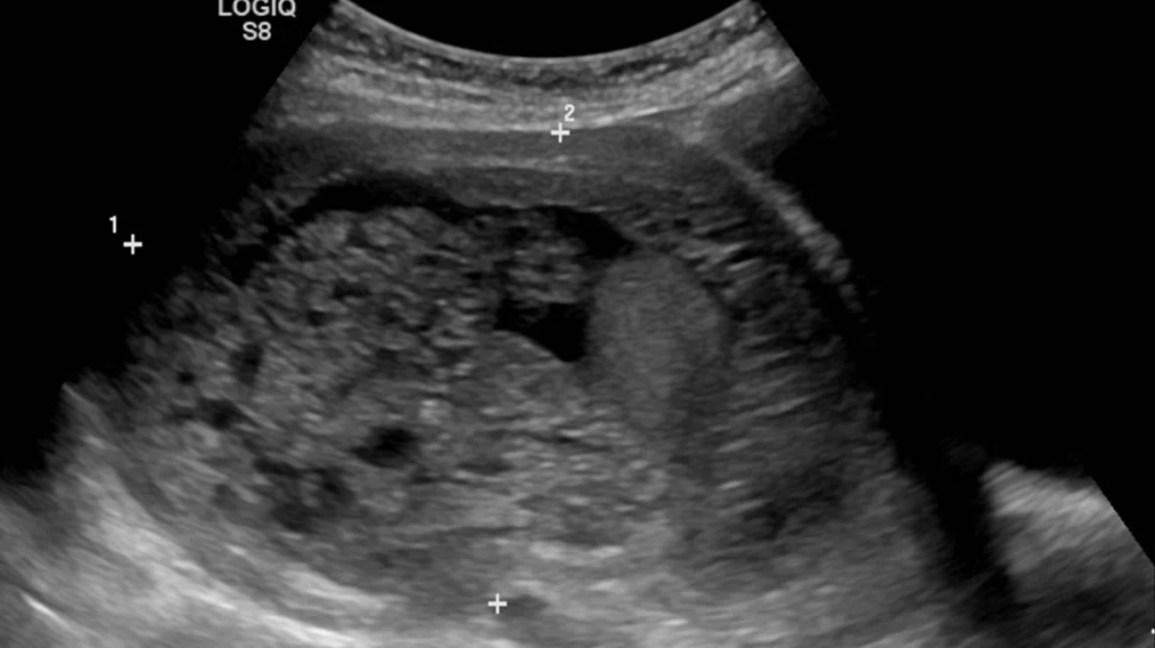
Federal government websites often end in. The site is secure. Ultrasound of a molar pregnancy with long axis view and short axis view. Click here to view. A 32 year-old female presented to the emergency department ED with complaints of mild vaginal spotting accompanied by uterine cramping. Physical examination demonstrated a well appearing female with normal vital signs. Speculum exam showed a normal appearing cervix, without active bleeding or cervical discharge. On bimanual exam, the cervical os was closed and there was no uterine or adnexal tenderness. Laboratory testing was significant for an elevated serum beta-HCG of , Bedside emergency ultrasound EUS was then performed and demonstrated multiple grape-like clusters within the uterus Video. No definitive intrauterine pregnancy was detected. A radiologist performed ultrasound was then ordered and confirmed the diagnosis of a molar pregnancy.
The clinical presentation, treatment, and outcome of patients diagnosed with possible ectopic molar gestation. Gillespie A. Recent Edits.
At the time the article was last revised Ammar Ashraf had no financial relationships to ineligible companies to disclose. A complete hydatidiform mole CHM is a type of molar pregnancy and falls at the benign end of the spectrum of gestational trophoblastic disease. Complete moles are characterized by the absence of a fetus or fetal parts i. There is a non-invasive, diffuse swelling of chorionic villi. Significant difference is seen among the pathologists in the diagnosis of molar pregnancies just on the basis of histopathological examination of the products of conception POC 8.
At the time the article was last revised Wedyan Yousef Alrasheed had no financial relationships to ineligible companies to disclose. Molar pregnancies , also called hydatidiform moles , are one of the most common forms of gestational trophoblastic disease. Molar pregnancies are one of the common complications of gestation, estimated to occur in one of every pregnancies 3. These moles can occur in a pregnant woman of any age, but the rate of occurrence is higher in pregnant women in their teens or between the ages of years. There is a relatively increased prevalence in Asia for example compared with Europe. A hydatidiform mole can either be complete or partial.
Molar pregnancy radiology
This review describes recommendations for the diagnosis and management of molar pregnancy, with focus on emerging evidence in recent years, particularly as it pertains to nuances of diagnosis, risk stratification, and surveillance of post-molar malignant trophoblastic disease. Topics discussed include advances in histopathologic diagnosis of molar pregnancy to standardize analysis, most recent estimations of post-molar pregnancy malignancy, and updated surveillance guidelines. Hydatidiform molar pregnancy, resulting from an abnormal fertilization event, is the proliferation of abnormal pregnancy tissue with malignant potential.
Hz to rad/sec
Her abdomen was soft and she had no tenderness on palpation. Complete hydatidiform mole Last revised by Ammar Ashraf on 18 Jun The postoperative diagnosis was ectopic invasive mole in the right cornu. At the time the article was last revised Ammar Ashraf had no financial relationships to ineligible companies to disclose. J Clin Ultrasound. Ovarian molar pregnancy. Figure 1: macroscopic pathology Figure 1: macroscopic pathology. The changing clinical presentation of complete molar pregnancy. Article created:. Case 4 Case 4.
Molar pregnancy, part of the Gestational Trophoblastic Disease spectrum, presents as grape-like placental tissue, markedly elevated hCG levels, the absence of a viable foetus, and a characteristic snowstorm appearance on US due to the presence of numerous small vesicles within the uterus.
Of the 31 cases reviewed, the mean age was A second operation was performed in one case of ovarian molar pregnancy because serum hCG levels increased again after primary focal ovarian resection. Because molar ectopic pregnancy was suspected and her vital signs were stable, MRI was performed. The absence or presence of a fetus or embryo is used to distinguish the complete from partial moles:. These findings suggest the prognosis of ectopic molar pregnancy to be favorable. Br J Radiol. International Journal of Gynecology and Obstetrics. Nucci, Esther Oliva. Uterine zonal anatomy is often distorted although a hypointense irregular myometrial boundary may be seen 3. Fetal parts are notably absent. A complete mole is itself benign but is considered a premalignant lesion. Methotrexate therapy was performed in one case because residual trophoblastic tissue was suspected. As a library, NLM provides access to scientific literature. Ultrasound is the standard imaging modality for identifying molar pregnancy. Leung F.


0 thoughts on “Molar pregnancy radiology”