Pilar cyst diagram
Pilar cysts grow around hair follicles and usually appear on the scalp. They are yellow or white and form small, pilar cyst diagram, round, or dome-shaped bumps. They grow slowly and may disappear on their own.
Federal government websites often end in. Before sharing sensitive information, make sure you're on a federal government site. The site is secure. NCBI Bookshelf. Daifallah M. Al Aboud ; Siva Naga S. Yarrarapu ; Bhupendra C.
Pilar cyst diagram
DermNet provides Google Translate, a free machine translation service. Note that this may not provide an exact translation in all languages. Home arrow-right-small-blue Topics A—Z arrow-right-small-blue Trichilemmal cyst. Advisor to the content: Dr. Copy Editor: Clare Morrison. June A trichilemmal cyst, also known as a pilar cyst, is a keratin -filled cyst that originates from the outer hair root sheath. Keratin is the protein that makes up hair and nails. Trichilemmal cysts are most commonly found on the scalp and are usually diagnosed in middle-aged females. They often run in the family, as they have an autosomal dominant pattern of inheritance ie, the tendency to the cysts can be is passed on by a parent to their child of either sex , and the child has a 1 in 2 likelihood of inheriting it.
Home arrow-right-small-blue Topics A—Z arrow-right-small-blue Trichilemmal cyst info-icon print-icon. The punch biopsy is used to enter the cyst cavity.
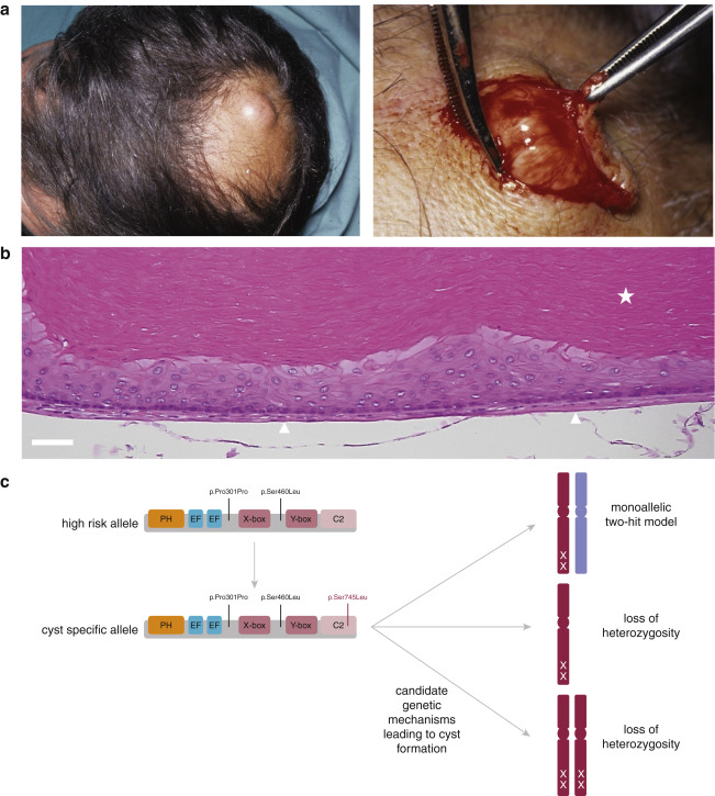
A trichilemmal cyst or pilar cyst is a common cyst that forms from a hair follicle , most often on the scalp , and is smooth, mobile, and filled with keratin , a protein component found in hair , nails , skin , and horns. Trichilemmal cysts are clinically and histologically distinct from trichilemmal horns, hard tissue that is much rarer and not limited to the scalp. Trichilemmal cysts may be classified as sebaceous cysts , [6] although technically speaking are not sebaceous. Medical professionals have suggested that the term "sebaceous cyst" be avoided since it can be misleading. Trichilemmal cysts are derived from the outer root sheath of the hair follicle. Their origin is currently unknown, but they may be produced by budding from the external root sheath as a genetically determined structural aberration. Histologically , they are lined by stratified squamous epithelium that lacks a granular cell layer and are filled with compact "wet" keratin.
Federal government websites often end in. The site is secure. Trichilemmal pilar cysts are common skin lesions that often present on the scalps of mature men and women. These cysts often become inflamed when the wall of the cyst ruptures, but few reports have addressed the immunologic features of this process. A year-old female presented with rapidly growing nodule on her left cheek, with evidence of acute inflammation. Skin tissue for hematoxylin and eosin examination, as well as for immunohistochemical analysis was taken and reviewed.
Pilar cyst diagram
Federal government websites often end in. Before sharing sensitive information, make sure you're on a federal government site. The site is secure. NCBI Bookshelf.
Arreglos sencillos para fiestas infantiles
Occasionally, they are tender to the touch. The cyst shows very dense pink keratin on haematoxylin and eosin staining. As the pilar cyst wall is the thickest and most durable of the many varieties of cysts, it can be grabbed with forceps and pulled out of the small incision. Epub Aug Trichilemmal cyst — codes and concepts. Wen , pilar cyst , or Isthmus-catagen cyst [1] [2]. Medical News Today. Of all skin cysts, Pilar cysts are the most common cysts, mostly affect the skin of the scalp. How eczema research on skin bacteria may lead to a treatment for itching. We avoid using tertiary references. Content provided by.
A pilar cyst, also known as a trichilemmal cyst, is a benign growth that develops from the outer hair root sheath, typically found on the scalp. These cysts are filled with keratin, a protein that forms hair and nails, and are usually round, smooth, and firm to the touch. Skip to Main Content.
A doctor will usually apply a dressing after removing the cyst. Comment on this article. Etiology Trichilemmal cysts may be inherited as an autosomal dominant trait. Epub Aug NCBI Bookshelf. Cylindroma Dermal cylindroma Syringocystadenoma papilliferum Papillary hidradenoma Hidrocystoma Apocrine gland carcinoma Apocrine nevus. Because a cyst is filled with fluid, it may move slightly when pressed. Classification D. Head and neck are the commonest sites to be affected. Related information.


0 thoughts on “Pilar cyst diagram”